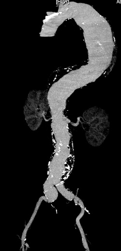
Angiogram showing a Thoracic Aneurysm


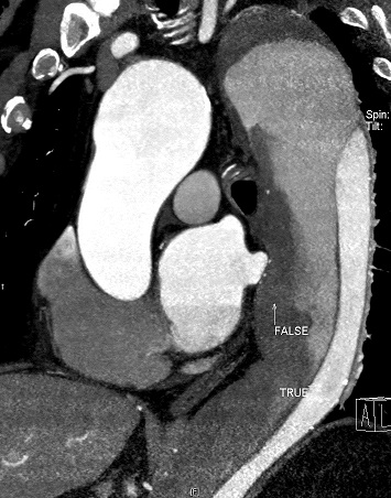
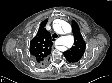
CT Angiograms showing a Thoracic Dissection
When an aneurysm occurs in the chest it is called a Thoracic aneurysm. The thoracic aorta can also develop a split within the layers of the artery wall, this is called a dissection. The risk factors for developing a Thoracic aneurysm are similar to those for an abdominal aneurysm. The congenital conditions with weaker aortic walls can often be associated with thoracic aneurysms and dissestions. Most thoracic aneurysms will be found by scanning or xrays, few present with symptoms. These are monitored when small in size, and are usually considered for intervention to treat the aneurysm when 6cms in size. As with AAA, the aim is to prevent rupture of the aneurysm. Treatment with stents has improved the results for thoracic aneurysms. When the aneurysm is near the heart, open surgery may be required. For extensive aneurysms where a lot of the aorta is dilated a combination of surgery and stenting, a "hybrid" procedure may be needed.
Dissection of the thoracic aorta is a cause of chest pain. This may suggest the diagnosis, but can be mistaken for heart attack, chest infection, lung embolus, and chest muscle or rib pain. A CT scan can show the dissection. If it is close to the heart, an urgent repair is needed, and that often involves surgery with heart bypass carried out by cardiothoracic surgeons.
Dissection further away from the heart can be monitored, with the blood pressure controllled at a low level. Scans will show if the aorta is stabilising. If not a stent can be used to help close the "split" or "tear" in the aorta. This is a newer treatment option and is still being evaluated, but is useful in some cases.
In some cases the aorta does stabilise, pain settles and it is safe to monitor it over time. Blood pressure control remains important.
For more information click HERE